-
November 13, 2025
-
5 min read
ETT Cuff Inflation: The Importance of Manometers
By Courtney Scales DipVN NCert (Anaesth) RVN
Airway devices are used to secure and maintain and protect a patent airway, provide a means to deliver oxygen and volatile gases, remove expired waste gases and support ventilation.
General anaesthesia inhibits protective airway reflexes such as coughing and swallowing, which normally prevent foreign material from entering the lower airway. The loss of these reflexes may put the patient at risk of aspirating foreign material e.g., gastroesophageal reflux (GOR) or fluid from the dental scaler. An endotracheal tube (ETT) remains the gold standard in airway protection1.
ETTs may be made from red rubber, polyvinylchloride (PVC) or silicone and can be manufactured with or without a polyurethane cuff. There are two different types of cuffs available:
- High Pressure/Low Volume (HP/LV) - A lower volume of air is required to inflate the cuff, but it creates a narrow area of high pressure on the mucosa. The cuff lays flat against ETT when deflated
- Low Pressure/High Volume (LP/HV) - A larger volume of air is required to inflate the cuff however the pressure is spread out throughout the length of the cuff, reducing damage to the mucosa. The cuff is wrinkled against the ETT when deflated
Once intubation is achieved and there is confirmation of correct placement, the ETT should be secured with a tie, before the inflation of the cuff. The cuff must be checked with only oxygen being delivered to the patient before the volatile agent is administered.
There are many different techniques to inflate the ETT cuff
A manometer accurately measures the pressure within the cuff against the mucosal wall, where the pressure should not exceed 25mmHg or 34cmH2O2. If the cuff is inflated to pressures above 35mmHg, then capillary perfusion may become compromised and injuries can occur, which can cause tracheal mucosal irritation, ischemia or necrosis2. The manometer may be attached to a manual pump (similar to a sphygmomanometer) or be a syringe-based manometer such as the AG Cuffill® and Tru-Cuff devices. The AG Cuffill® offers a higher percentage of properly inflated cuffs (86.7%) compared to the Tru-Cuff (50%)3.
The Minimal Occlusion Volume (MOV) technique is the most popular in practice however only 3.3% of cuffs inflated with the MOV technique have been reported to be at a satisfactory pressure4. This technique involves one person delivering a positive pressure breath to the patient by inflating the lungs to 16-18cmH2O and the second person inflating the cuff until no audible leak is heard5.
When using palpation of the pilot balloon and MOV for cuff inflation, optimal intracuff pressures are rarely reached despite no leaks being heard around the cuff when providing PPV. As experience does not affect this skill, a cuff manometer is recommended5.
It is important to note that the cuff of the ETT may increase in size when nitrous oxide is being used1.
An overinflated cuff can be just as dangerous to the patient as an underinflated cuff, which puts the patient at risk of aspirating foreign material and exposes theatre personnel to anaesthetic gases.
In cats, longitudinal tears form along the length of the trachealis muscle due to trauma when using ETTs, with most tears approximately 2-5cm in length6. There is no difference in the incidence of occurrence of a tracheal tear when using either HP/LV or LP/HV cuffs, it is only the length of injury that differs, with overinflated cuffs being a contributing factor7.
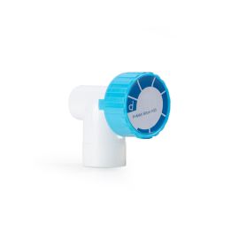
The AG Cuffill
The AG Cuffill is a cuff inflator and manometer, suitable for use with several airway devices: ETTs, tracheostomy tubes and laryngeal mask airways, including supraglottic airway devices. It isn’t suitable for checking the pressure of liquid-based balloons, such as those on foley urinary catheters.
It will inflate and monitor the cuff pressure of the airway device between the ranges of 0-99cmH2O, with an accuracy of ±2cmH2O and is designed to be used up to 100 times. The remaining uses blink onto the display before displaying “00”, indicating it is ready for use.
More display codes are detailed in the package insert, as are the cleaning and disinfection instructions.
To inflate the cuff:
- Press the yellow power button to the right of the display. It should blink twice and display “00”
- Pull back on the plunger
- Connect it to the airway device’s cuff inflation line
- Inflate the cuff until the desired pressure is reached
- If the required pressure is still not achieved (e.g., large volume cuffs) then disconnect, and repeat steps 2 to 4
- Disconnect the AG Cuffill from the airway device. Please note, this may cause a pressure drop by 1-2cmH2O
To check the cuff pressure:
- Press the yellow power button to the right of the display. It should blink twice and display “00”
- Push the plunger in until it stops
- Connect it to the airway device’s cuff inflation line and read the pressure
- Inflate or deflate the cuff until the desired pressure is reached
- Disconnect the AG Cuffill from the airway device. Please note, this may cause a pressure drop by 1-2cmH2O
References
1. Hughes L. Breathing systems and ancillary equipment. In: Duke-Novakovski T, de Vries M, Seymour C. BSAVA Manual of Canine and Feline Anaesthesia and Analgesia. 3rd ed. Gloucester: British Small Animal Veterinary Association (BSAVA); 2016.
2. Hartsfield S. Airway management and ventilation. In: Tranquilli W, Thurman J, Grimm K, ed. by. Lumb and Jones Veterinary Anesthesia and Analgesia. 4th ed. Oxford: Blackwell; 2007. p. 495-512.
3. Hung W, Ko J, Weil A, Weng H. Evaluation of Endotracheal Tube Cuff Pressure and the Use of Three Cuff Inflation Syringe Devices in Dogs. Frontiers in Veterinary Science. 2020;7.
4. White D, Redondo J, Mair A, Martinez-Taboada F. The effect of user experience and inflation technique on endotracheal tube cuff pressure using a feline airway simulator. Veterinary Anaesthesia and Analgesia. 2017;44(5):1076-1084.
5. Robertson S, Gogolski S, Pascoe P, Shafford H, Sager J, Griffenhagen G. AAFP Feline Anesthesia Guidelines. Journal of Feline Medicine and Surgery. 2018;20(7):602-634.
6. Mitchell S, McCarthy R, Rudloff E, Pernell R. Tracheal rupture associated with intubation in cats: 20 cases (1996–1998). Journal of the American Veterinary Medical Association. 2000;216(10):1592-1595.
7. Quandt J. Postintubation Tracheal Tears in Cats [Internet]. Clinicals Brief. 2017 [cited 2 August 2022]. Available from: https://www.cliniciansbrief.com/article/postintubation-tracheal-tears-cats
Share on Facebook
Share on X
Share on Pinterest
Share on LinkedIn
Related Products
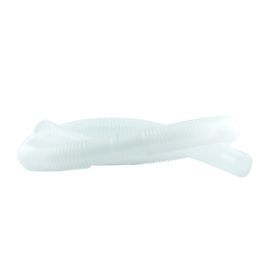
Gas Scavenging Tubing
€8.72
€7.09
APL Valve 10cm
€13.00
€13.00
Reservoir Bag
€8.54
€6.94
Vetronic Merlin Small Animal Ventilator
€8,175.92
€6,647.09

Popular Posts